Please wait...

Please wait...
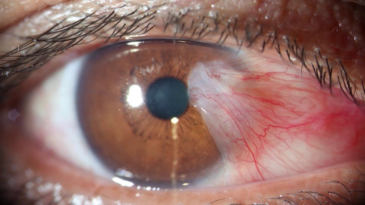
The pterygium consists of an abnormal growth of triangular-shaped tissue that extends from the conjunctiva (transparent membrane that covers the sclera, the white part of the eye) to the cornea (anterior and transparent surface of the eye).
The pterygium consists of an abnormal growth of triangular-shaped tissue that extends from the conjunctiva (transparent membrane that covers the sclera, the white part of the eye) to the cornea (anterior and transparent surface of the eye). It manifests as a kind of whitish «cloth» on the inner and / or outer edge of the cornea.
At first, the pterygium or fleshyness in the eye can be painless although the symptoms it causes usually depend on the size it acquires. As the tissue grows, it is usual to produce a sensation of a foreign body, burning, red eye, tearing and, even, it can prevent vision, hinder flickering or induce the appearance of astigmatism. In these cases, the ophthalmologist usually recommends pterygium surgery. On some occasions, the pterygium can be confused with the pinguecula, which is a benign accumulation of protein and fat in the form of rice grain that appears on the conjunctiva, and not on the cornea, such as the pterygium.
The exact cause of the appearance of the pterygium has not been fully defined, although it is usually more frequent in people who suffer from dry eyes or who spend a lot of time outdoors under sun exposure. Therefore, by adopting certain behaviors, such as protecting ourselves from the sun (ultraviolet light) or combating dry eyes, we can prevent their appearance or develop more quickly and even make vision difficult.
The treatment of the pterygium will depend on how the tissue in the eye (or the eyes) evolves, on the speed at which it grows and on the phase in which it is found.
When the pterygium is incipient or very small, ophthalmologists often use steroids to reduce inflammation and lubricating drops to lessen the sensation of a foreign body in the eye.
If the pterygium reaches a size that compromises vision reaching the pupillary area or becomes especially unsightly, the ophthalmologist may consider removing it by surgery.
Pterygium surgery should be performed by an ophthalmologist specializing in ocular surface surgical techniques. In recent years, conjunctive-free autograft surgery has become very common in ophthalmology, which is that, while the surgeon removes the pterygium, it places a small portion of the patient’s own conjunctiva at the site where previously removed the tissue. This conjunctiva portion can be fixed by absorbable sutures or by tissue adhesives (biological glues). Pterygium surgery is usually performed with local anesthesia and without admission, it is an outpatient treatment.
Even applying surgical techniques, there is a possibility that the pterygium reappears, especially if it was very evolved and already invaded the pupillary area. Therefore, its level of recurrence is quite high.
